Imaging Flow Cytometry and Flow Cytometric Analysis Core
- Department of Cell and Molecular Biology Home
- About the Department
- Education
- Research
- Contact Cell and Molecular Biology
- Support the Department of Cell and Molecular Biology
- Support the Center for Immunology and Microbial Research and the Graduate Program in Microbiology and Immunology
Imaging Flow Cytometry and Flow Cytometric Analysis Core
Purpose
The Imaging Flow Cytometry and Flow Cytometric Analysis Core provides the UMMC research community with expertise, equipment access, and assistance with experimental design and data analysis.

Equipment
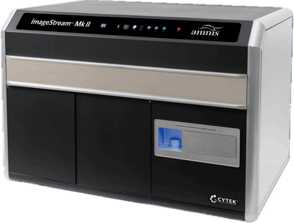
Amnis ImageStreamX MkII
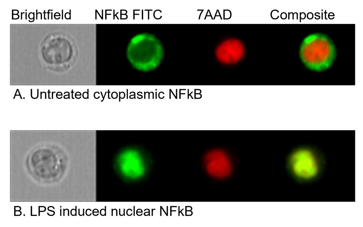
The Amnis ImageStreamX MkII is a benchtop imaging flow cytometer system that combines the detailed cellular images and morphologic information of microscopy with the large sample size and phenotyping abilities of flow cytometry. The system has two CCD cameras, three lasers (Violet 405 nm, Blue 488 nm, Red 642 nm), and three imaging objectives (20x, 40x, 60x). The ImageStreamX MkII captures multiple images of each cell in flow, including brightfield, darkfield, and up to 10 fluorescent markers simultaneously. The image resolution depends on the magnification settings, with pixel sizes of 1 μm2, 0.25 μm2, and 0.1 μm2, respectively.

Data analysis is performed using the IDEAS software, which utilizes wizard-based and customized workflows to objectively quantify distinct cell populations, cellular features, and processes, including nuclear translocation, shape change, internalization, and apoptosis.
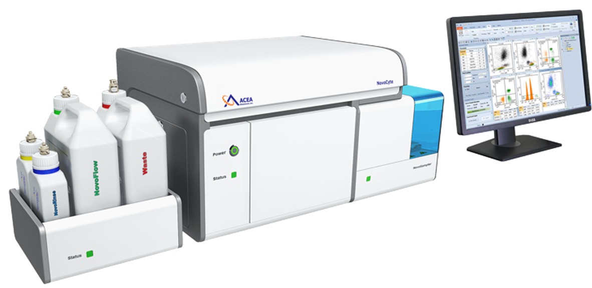
NovoCyte 3000 Flow Cytometer
 The NovoCyte 3000 is a high throughput flow cytometric analyzer with three lasers (Violet 405 nm, Blue 488 nm, Red 642 nm) and filters for detecting up to 13 fluorescent colors. The instrument is equipped with the NovoSampler Pro multi-sampler that accommodates 24-, 48-, or 96-well plates or a 24-tube rack. The Novocyte uses the NovoExpress software for data acquisition and analysis. Data files can be exported as FCS 2.0 or FCS 3.1.
Personnel
- Director: Eva Bengtén, PhD,
Professor, Department of Cell & Molecular Biology
Office G562
Location
- The Amnis ImageStreamX MkII is in room G669, a BSL-1 designated laboratory, and the NovoCyte 3000 flow cytometer is in the BSL-2 laboratory, G667, in the Guyton Research Building.
Core services
The core provides training in imaging flow cytometry, flow cytometric analysis, and consultations on experimental design and data interpretation. The core is not a drop-off service facility. After being trained and certified, the user operates the instruments.
To discuss an experiment or book the instrument, please contact Dr. Eva Bengtén.
IMPORTANT: Because the Amnis ImageStreamX MkII was purchased using NIH funds, (Mississippi Center of Excellence in Perinatal Research (MS-CEPR)-COBRE, Award: P20GM121334), publications utilizing data acquired with the ImageStream must acknowledge this grant. Please use the following language in publications in which you have used the ImageStream for your experiments:
"Research reported in this publication was supported by the National Institute of General Medical Sciences of the National Institutes of Health under Award Number P20GM121334. The content is solely the responsibility of the authors and does not necessarily represent the official views of the National Institutes of Health."


